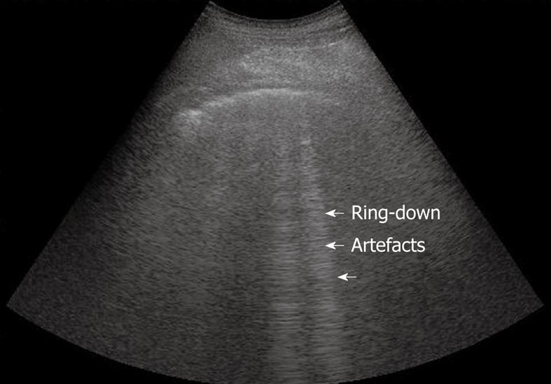
Yet clinical experience and review of the literature show that lung ultrasound has been previously proposed for diagnosing pneumothorax ( 1-4) or alveolar consolidation ( 5-8). The lung is therefore usually considered poorly accessible to ultrasound. Can ultrasound, a noninvasive, easily implemented technique, be of any use? Basically, the problem is that air stops the progression of the ultrasound beam, and only reverberation artifacts are visible under the lung surface. However, in critical care units, chest X-ray is performed at the bedside and technologic deficiencies may make this diagnosis difficult. The diagnosis of alveolar-interstitial syndrome is based on chest X-ray. In conclusion, presence of the comet-tail artifact allowed diagnosis of alveolar-interstitial syndrome. Tomodensitometric correlations showed that the thickened sub-pleural interlobular septa, as well as ground-glass areas, two lesions present in acute pulmonary edema, were associated with the presence of the comet-tail artifact. It was absent or confined to the last lateral intercostal space in 120 of 129 patients with normal chest X-ray (specificity of 93.0%). This pattern was present all over the lung surface in 86 of 92 patients with diffuse alveolar-interstitial syndrome (sensitivity of 93.4%). The ultrasonic feature of multiple comet-tail artifacts fanning out from the lung surface was investigated. The antero-lateral chest wall was examined using ultrasound. 8.6 163-166.Can ultrasound be of any help in the diagnosis of alveolar-interstitial syndrome? In a prospective study, we examined 250 consecutive patients in a medical intensive care unit: 121 patients with radiologic alveolar-interstitial syndrome (disseminated to the whole lung, n = 92 localized, n = 29) and 129 patients without radiologic evidence of alveolar-interstitial syndrome. More research is needed to know whether this finding is clinically relevant, but the prospects are looking bright. However, visualizing abdominal A-lines in every quadrant is concerning for bowel perforation in the cases presented. It is unclear what role the identification of abdominal A-lines can play in patient management, as this can be a normal finding when loops of gas-filled bowel are adherent to the abdominal peritoneum. However, explicit knowledge of sonographic artifacts such as the reverberation artifact can identify unexpected emergent diagnoses. The sensitivity of ultrasound for these diagnoses will likely never be adequate to be used as a screening tool. Point-of-care ultrasound is not the test of choice to rule out necrotizing fasciitis, all foreign bodies, or bowel perforation. The patient is diagnosed with emphysemtaous cholecystitis and subsequently undergoes an uncomplicated cholecystectomy.

A bedside ultrasound is completed as shown (Image 5).

She appears well, without jaundice, but has a fever and a positive Murphy's sign. Gallbladder with several reverberation artifacts originating within the gallbladder wall.Ī 48-year-old female presents for right upper quadrant abdominal pain. A diagnosis of necrotizing fasciitis is made, and the patient is taken to the operating room for debridement.
#Comet tail artifact free
The radiograph confirms free air without retained foreign body.

The ultrasound (Image 1) demonstrates an impressive reverberation, ring-down artifact, concerning for a retained metal foreign body versus free air. A bedside ultrasound is performed, followed by a plain radiograph. His right arm demonstrates evidence of significant cellulitis and possible abscess near the antecubital fossa. You suspect the patient is intoxicated from methamphetamines. 1Ī-lines are part of the normal sonographic thoracic exam, but reverberation artifacts in other anatomic regions can be pathologic and point toward life-threatening diagnoses.Ī 38-year-old male presents with worsening arm pain and swelling. Another example of reverberation artifact is known as ring-down artifact and typically originates from small metal objects or air bubbles. Comet tail artifacts are another type of reverberation artifact originating from the pleural surface.

A-lines are the reverberated ultrasound waves that have bounced between the parietal and visceral pleura. A common example is the “A-lines” visualized on thoracic ultrasound. Reverberation artifacts occur when two interfaces with high acoustic impedance bounce the ultrasound waves between them. Reverberation artifact originating from the patient's upper extremity soft tissue.


 0 kommentar(er)
